


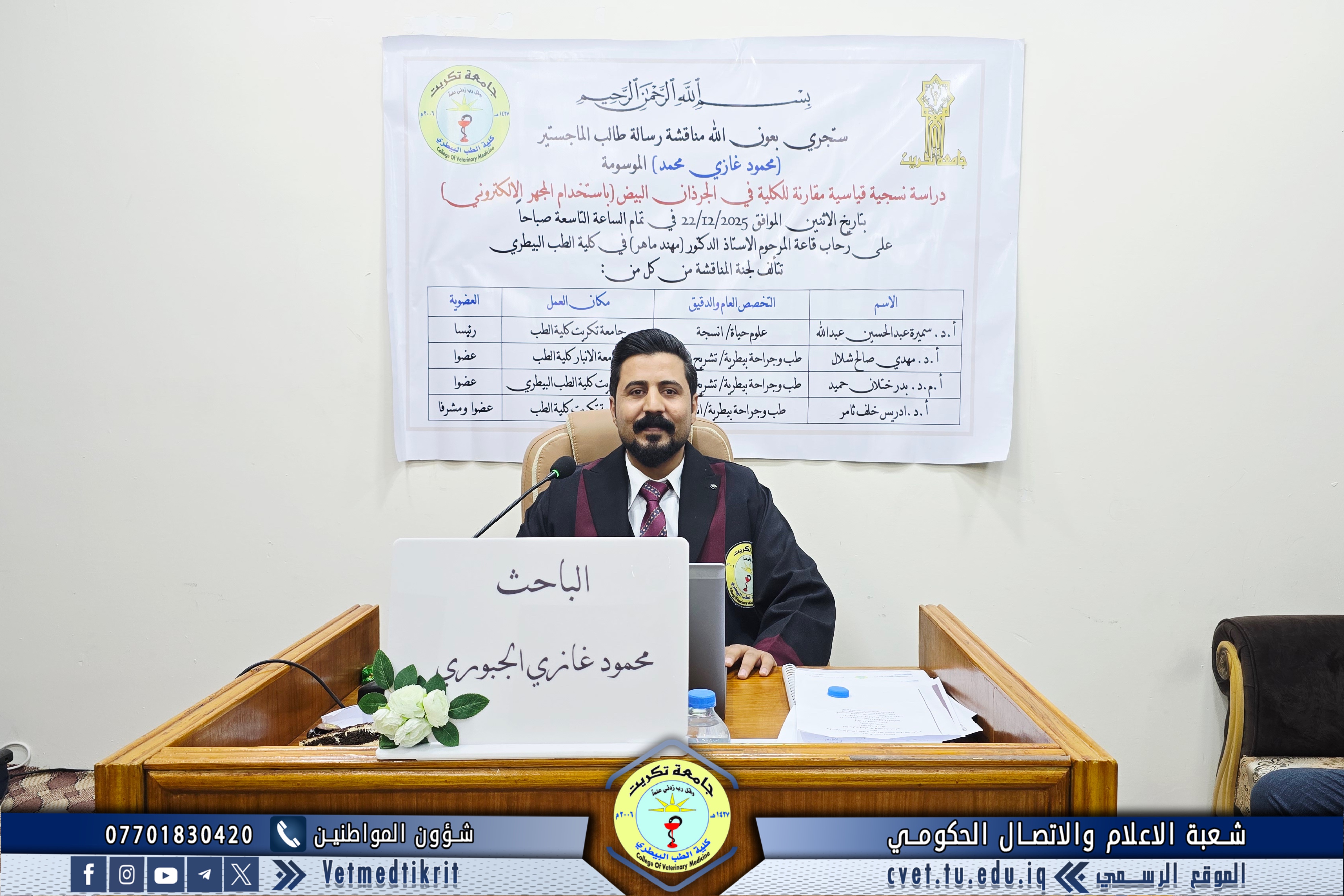


By the grace of Almighty God, the College of Veterinary Medicine at the University of Tikrit held the public defense of the Master’s thesis entitled “A Comparative Standardized Histological Study of the Kidney in Albino Rats (Using the Electron Microscope)”, submitted by the student Mahmoud Ghazi Mohammed in the field of Veterinary Histology.
The examination committee consisted of the following members:
-
Professor Dr. Samira Abdulhussein Abdullah
Specialization: Histology
University of Tikrit – College of Medicine
Chairperson -
Professor Dr. Mahdi Saleh Shalal
Specialization: Anatomy and Histology
University of Anbar – College of Medicine
Member -
Assistant Professor Dr. Badr Khatlan Hamid
Specialization: Anatomy and Histology
University of Tikrit – College of Veterinary Medicine
Member -
Professor Dr. Idris Khalaf Thamer
Specialization: Histology
University of Tikrit – College of Medicine
Member and Supervisor
Abstract of the Thesis
The present study was conducted at the College of Veterinary Medicine / University of Tikrit, in the animal house affiliated with the college, during the period from August 2024 until the end of December 2024. Laboratory animals consisting of male albino rats were used, with a total number of 18 rats, divided into three groups, six rats per group.
The first group included rats aged two months, the second group included rats aged four months, and the third group included adult rats aged six months.
After housing the animals according to the experimental groups, dissection was performed by anesthetizing the animals and opening the thoracic region (sternum) downward to the pelvic region to extract the target organs of the study, namely the kidneys. Part of the samples was used for gross examination, while another part was placed in plastic tubes containing 10% formalin for sectioning and staining for histological examination. Additional kidney samples were preserved for electron microscopic studies.
The results of the study demonstrated significant differences among the kidneys of the three groups, with statistically significant differences in the measurements used in the study. The thickness of the renal cortex was 245.00 ± 2.54 at 30 days, 267.0 ± 2.32 at 90 days, and 287.00 ± 2.33 at 120 days. The thickness of the renal medulla was 299.11 ± 2.98 at 30 days, 303 ± 4.43 at 90 days, and 335.00 ± 2.33 at 180 days.
The standardized histological study showed differences in the number of mesangial cells within the glomeruli, with mean values for the three age groups (respectively) of 7.36 ± 0.02, 6.46 ± 0.01, and 5.45 ± 0.00.
Histological measurements of glomerular diameters also showed mean values of 47.31 ± 0.11, 51.74 ± 0.13, and 57.17 ± 0.06 for the three age groups, respectively.
Measurements of glomerular and Bowman’s capsule diameters were 34.66 ± 0.12, 42.29 ± 0.16, and 48.74 ± 0.16, while Bowman’s capsule measurements were 12.65 ± 0.06, 9.45 ± 0.08, and 8.43 ± 0.06, respectively.
The study also recorded measurements of renal tubule diameters. The mean values for the proximal tubules were 27.32 ± 0.08, 27.47 ± 0.12, and 27.92 ± 0.12, while the distal tubules measured 22.53 ± 0.07, 22.12 ± 0.10, and 22.25 ± 0.11 for the three rat age groups, respectively.
Measurements of the collecting tubules showed mean values of 30.62 ± 0.04, 30.72 ± 0.08, and 31.46 ± 0.04, with statistically significant differences among the age groups.
Electron microscopic analysis of glomerular measurements in rats aged two, four, and six months revealed substantial age-related increases in all dimensions, peaking with a dramatic 10.8-fold increase in glomerular volume between two and six months. This growth pattern represents normal postnatal renal maturation in rats and has important implications for experimental design, data interpretation, and applied research. The most notable finding was the continued rapid growth between four and six months of age, indicating that even at six months, rat glomeruli had not yet reached their maximum size. This contrasts with human kidney development, where glomerular size stabilizes early in adulthood.
The study confirmed that analysis of glomerular measurements across different age groups (two, four, and six months) in rats reveals clear age-related changes in glomerular dimensions and volume. These findings place the results within a broader understanding of kidney development, physiological function, and their potential implications for research and clinical applications.
The thesis defense, held in Dr. Muhannad Maher Hall at the College of Veterinary Medicine, was attended by a number of faculty members and a group of students.